Understanding the brain’s electrical activity during rest can unlock insights into how the body heals, restores energy, and forms memories. EEG sleep technology provides a precise window into these processes, using sensors to reveal brainwave patterns that define each stage of the nightly sleep cycle. By examining REM sleep EEG characteristics and the broader sleep stages EEG framework, researchers and clinicians gain valuable details to improve sleep health and diagnose disorders.
Monitoring the brain’s shifting activity overnight offers more than just academic knowledge. The ability to distinguish between sleep stages supports targeted interventions for insomnia, narcolepsy, and other sleep-related conditions. For anyone interested in optimizing rest, a closer look at EEG sleep reveals how subtle changes in brainwaves reflect the quality and depth of sleep, with practical implications for overall well-being.
What Is EEG Sleep and How Does It Work?
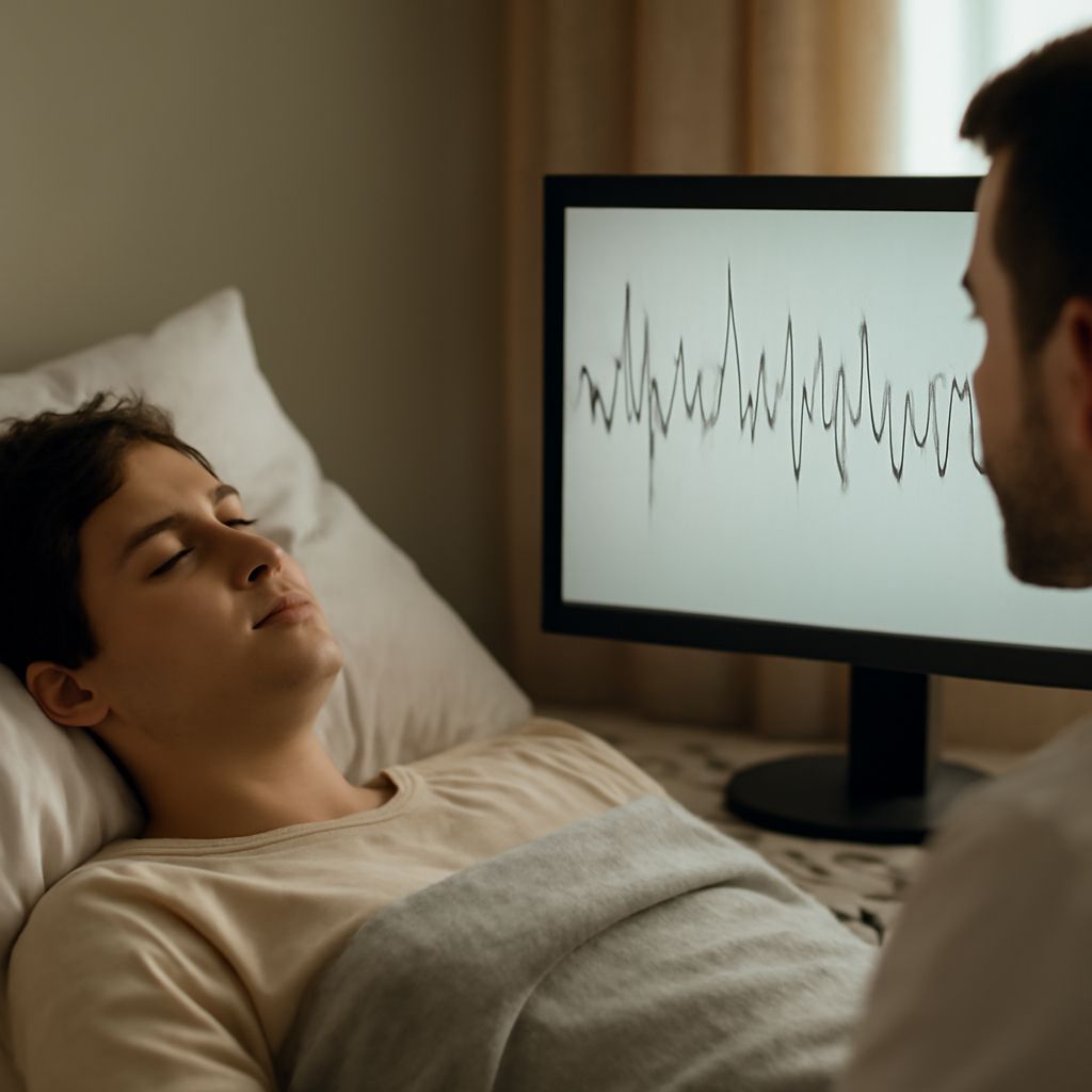
EEG sleep refers to the use of electroencephalography to monitor and record the brain’s electrical signals throughout the night. By placing small, strategically positioned electrodes on the scalp, clinicians can detect voltage fluctuations resulting from neurons firing in synchronized rhythms. These patterns, called brainwaves, change as a person drifts from wakefulness through different stages of sleep.
An EEG sleep procedure is non-invasive, painless, and commonly performed in sleep clinics as part of a polysomnography study. Electrodes are typically spaced according to the International 10-20 System, ensuring comprehensive coverage of the cerebral cortex. Each electrode picks up faint electrical signals, which are then amplified and charted as wavy lines—a real-time visual record of the brain’s activity.
Throughout the night, sleep cycles repeat in approximately 90-minute intervals, with the EEG capturing distinct shifts in frequency and amplitude. Each sleep stage—from light NREM sleep to deep slow-wave sleep and finally REM—has a unique brainwave signature. By analyzing these EEG traces, clinicians can identify abnormal patterns, such as disrupted cycles or unusual arousals, which may signal underlying sleep disorders.
The precise, continuous nature of EEG sleep measurement makes it the gold standard for research and diagnosis. Its ability to split sleep architecture into quantifiable segments allows for detailed sleep scoring, which in turn guides personalized treatment plans and informs sleep hygiene recommendations.
How EEG Tracks Sleep Stages: A Closer Look
EEG technology captures the dynamic progression through sleep, distinguishing between NREM (non-rapid eye movement) and REM (rapid eye movement) stages by examining subtle shifts in brainwave activity. During stage N1, the transition from wakefulness, the EEG shows a reduction in alpha waves and the emergence of low-voltage theta activity. This stage is fleeting, often lasting just a few minutes, yet it plays a crucial role in the sleep onset process.
As the sleeper enters stage N2, the EEG records hallmark features such as sleep spindles and K-complexes—brief bursts of activity and sharp waveform events, respectively, that indicate the brain is blocking external stimuli. This stage constitutes about half of a typical night’s sleep, providing stability and insulation from waking.
Deep sleep, or stage N3 (slow-wave sleep), is characterized by the dominance of high-amplitude, low-frequency delta waves. The EEG at this stage reflects synchronized neuronal firing across large brain regions, representing the most restorative phase for physical repair and immune function. Analyzing the proportion of time spent in N3 can help identify disorders such as sleep apnea or chronic sleep deprivation.
Finally, the transition into REM is marked by a dramatic change in the EEG pattern, with mixed-frequency, low-amplitude activity that resembles wakefulness. Only by segmenting and scrutinizing these recordings can the complex architecture of the human sleep cycle be fully understood and evaluated.
REM Sleep EEG: Unique Patterns and Their Meaning
REM sleep EEG analysis is central to decoding the mysteries of dreaming and memory processing. During this stage, the EEG reveals fast, irregular, low-voltage brainwave patterns, often mixed with sawtooth waves that are unique to REM. Unlike the synchronized delta rhythms of deep sleep, REM sleep EEG shows desynchronized activity—a sign of heightened brain activity even while the body remains almost completely still due to muscle atonia.
This paradoxical state, where the brain is active but the skeletal muscles are temporarily paralyzed, is easily distinguished in the REM sleep EEG recording. Such characteristics enable sleep technologists and neurologists to identify the precise onset and duration of REM periods, which typically increase in length over the course of the night, with the final REM episode lasting up to 40 minutes or more.
Changes in REM sleep EEG can offer clues about neurological health. For instance, decreases in REM density or abnormal intrusions of REM-like patterns into wakefulness may indicate narcolepsy or other central nervous system issues. By quantifying these patterns, clinicians can assess cognitive health, emotional regulation, and the effectiveness of certain medications or therapies.
Careful interpretation of REM sleep EEG provides a foundation for understanding how the brain processes daytime experiences, solidifies memories, and contributes to emotional balance. It is a critical component of comprehensive sleep analysis and research.
Why Understanding Sleep Stages EEG Matters for Health
Accurate measurement of sleep stages EEG supports early detection and management of a range of health conditions. For example, a shortened or fragmented slow-wave sleep phase, as revealed through EEG sleep studies, has been linked to higher risks of metabolic syndrome, cardiovascular disease, and impaired immune response. Similarly, tracking the balance between REM and NREM sleep can inform treatment strategies for depression, PTSD, and neurodegenerative disorders.
Sleep stages EEG is also vital for tailoring interventions in pediatric and geriatric populations, where age-specific changes in sleep architecture may impact growth, cognitive development, or risk of falls. For shift workers, athletes, or people recovering from illness, periodic EEG sleep assessments can help optimize recovery by pinpointing when sleep quality dips below the threshold needed for full body and mind restoration.
Beyond clinical applications, understanding EEG sleep patterns empowers individuals to make evidence-based adjustments to their nightly routines. Exposure to bright light, exercise, caffeine intake, and even stress management techniques can be tested for their impact on measurable stages of sleep. The objective nature of EEG data offers a clear benchmark for improvement, helping individuals set realistic goals and track progress over weeks or months.
Ultimately, integrating sleep stages EEG insights into public health and personal care underscores the essential role of sleep in long-term wellness, prevention, and performance.
Getting an EEG Sleep Study: What to Expect
Undergoing an EEG sleep study—commonly conducted as part of an overnight polysomnography—typically begins with a detailed intake assessment. Patients are asked to follow their normal routine before arriving at the sleep center in the evening. A sleep technologist will attach between 16 and 25 electrodes to the scalp, forehead, and sometimes additional sites on the face and chest, using conductive paste for stable readings.
Throughout the night, the EEG system records brainwave activity alongside other physiological signals such as eye movements (EOG), muscle tone (EMG), heart rate (ECG), and breathing patterns. Environmental controls, such as temperature and light, are carefully maintained to mimic the home sleep environment as closely as possible. The setup ensures that any disruptions to normal sleep are minimized, allowing for accurate staging and scoring.
After the study, a trained sleep specialist will analyze the EEG sleep data in 30-second epochs, assigning each segment to the appropriate stage based on established scoring manuals. The resulting hypnogram—a graphical representation of sleep stages over time—serves as the basis for diagnosis and treatment planning. Patients typically receive results within a week, along with tailored recommendations for improving sleep hygiene or addressing identified disorders.
EEG sleep studies set the standard for reliability and granularity in sleep assessment. Whether investigating unexplained daytime fatigue, suspected sleep apnea, or chronic insomnia, this approach gives both clinicians and patients actionable information to enhance sleep quality and overall health.